


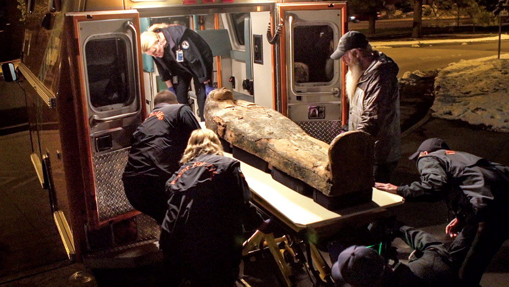
Flight for Life and DMNS staff transport an Egyptian mummy to Children’s Hospital.
In a time-bridging partnership, the Denver Museum of Nature & Science teamed up with Children’s Hospital Colorado to scan two mummies and a sarcophagus.
“The mummy transport is a unique example of our two worlds coming together and combining history with state-of-the art technology and medical expertise,” says Christy Dobson of Children’s Hospital Colorado.
The scans of two mummies and a sarcophagus were inspired by the Chicago Field Museum’s re-scanning of its mummies in 2011 and the valuable results that endeavor yielded. Denver’s 1991 scan of its mummies led museum scientists to believe one mummy had been relatively wealthy during her lifetime—and that served as the foundation for the “rich Mummy/poor mummy” storyline in the current exhibition. The latest scans suggest a different interpretation.
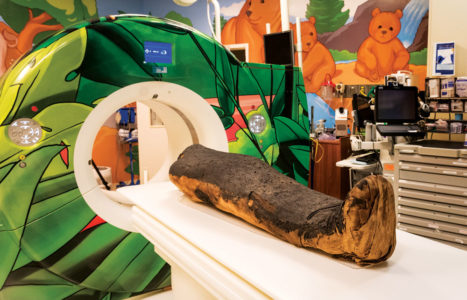
The mummy is pictured next to the Children’s Hospital scanner.
“Part of rescanning the mummies was because the rich mummy, poor mummy narrative is inaccurate. People had to have money to be mummified, so by definition there is no such thing as a poor mummy. It is true that the one mummy was not as carefully wrapped or buried with amulets and metal inside the wrappings, but she was placed in a gorgeously painted sarcophagus and the fact that she was mummified at all signals that her family cared for her and gave her the best treatment they could afford. We will be updating the narrative when we update the gallery with the results of the scientific analysis next year,” says Dr. Michelle Koons, curator of archaeology at the Museum of Nature & Science.
The hospital donated its expertise and equipment to allow the museum to perform CT scans, which use a series of X-ray images, to provide high-resolution pictures that help further enhance the understanding of the relics.
Flight for Life provided a de-commissioned ambulance and gurney for the scans. At the museum, the gurney was retrofitted with a platform that could hold and transport the mummies and sarcophagi. Starting in the evening after rush hour, each mummy or sarcophagus was loaded into the ambulance by museum and Flight for Life personnel and driven to the hospital, scanned and returned. Altogether, the process took about eight hours.
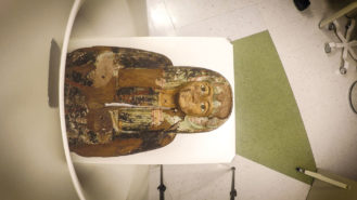
The sarcophagus is pictured partially in the scanner.
The mummies had been scanned before at University Hospital in 1991, and a single mummy was scanned again in 1998, but thanks to advances in technology, these most recent photographs taken on April 18, 2016, provide even more detailed imagery, increasing the potential for new insight and understanding of the ancient treasures. The first scan revealed astounding images within seconds. While it will take time to review and interpret the images, scientists at the scene were excited by the clarity and detail they showed.
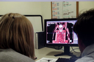
Scientists get a first look at what the scan reveals.
“The scans back in the ’90s were done with one-centimeter resolution,” explains Koons. “These new scans are at .5 mm resolution. We can see many more details of the wrapping technique and the amulets and metal pieces inside the wrappings. In fact, we can even see that the metal piece that is covering the incision where the organs were removed from mummy 1997-24.1. We are processing the data and hope to have images of this in the near future. We also hope to see if any pathologies, such as arthritis, can be identified on the bones.”
“Our ongoing partnership allows us to engage in our passion for science, medicine, the human body, nutrition, exercise and the overall well-being of our community,” says Dobson.
The full analysis of the CT scans and other tests are expected to be complete by the end of the year. In addition to the CT scans, several other minimally invasive tests will be done to further scientific understanding while protecting the integrity of the artifacts, including: radiocarbon dating of very small samples of the linens, skin, and wood from the sarcophagi; isotope analysis of the linen, hair and skin; core sampling of the sarcophagi for use in tree ring dating and wood analysis; and portable X-ray fluorescence analysis to determine the composition of the pigments on the sarcophagi.
“We look forward to adding the next chapter to its history when we get all the new data back in late fall,” says Koons. “This is an incredible opportunity for the museum and for the field of archaeology. A scientist may not get such an opportunity during her career. It’s been a privilege to work with such a dedicated team of medical and museum professionals.”

0 Comments